Most women are sent for one with almost no explanation, and the imagination fills the gap. So here is the plain version: what the scan does, what it feels like, what the numbers mean, and — most importantly — what a normal result does and does not close off.
Why has my doctor ordered a transvaginal ultrasound?
It is the workhorse of gynaecology at midlife. The usual reasons:
- Any bleeding after menopause. This is the big one. Bleeding after 12 consecutive months without a period is never normal and always needs investigation, even if it was one episode, even if it was a single streak on the paper. Most causes turn out to be benign — a thin, fragile atrophic lining is the commonest — but that is never assumed until it has been checked: around 1 in 10 women with postmenopausal bleeding has endometrial cancer, and it is one of the few cancers that usually announces itself early enough to be curable.
- Heavy, prolonged or erratic bleeding in perimenopause — to look for fibroids, polyps, adenomyosis or a thickened lining. (See also heavy periods and spotting between periods.)
- Pelvic pain or pressure, a swollen abdomen, or pain during sex.
- A lump or fullness felt on pelvic examination.
- Suspected ovarian cyst — to describe it (simple, complex, solid areas, blood flow) and decide whether it can simply be watched.
- Watery, pink or blood-stained vaginal discharge after menopause — which is treated with the same seriousness as frank bleeding.
- Checking an IUD's position, investigating fertility, or assessing early pregnancy.
What does a transvaginal ultrasound actually feel like?
Fear of the unknown is why women postpone this scan, so no coyness here.
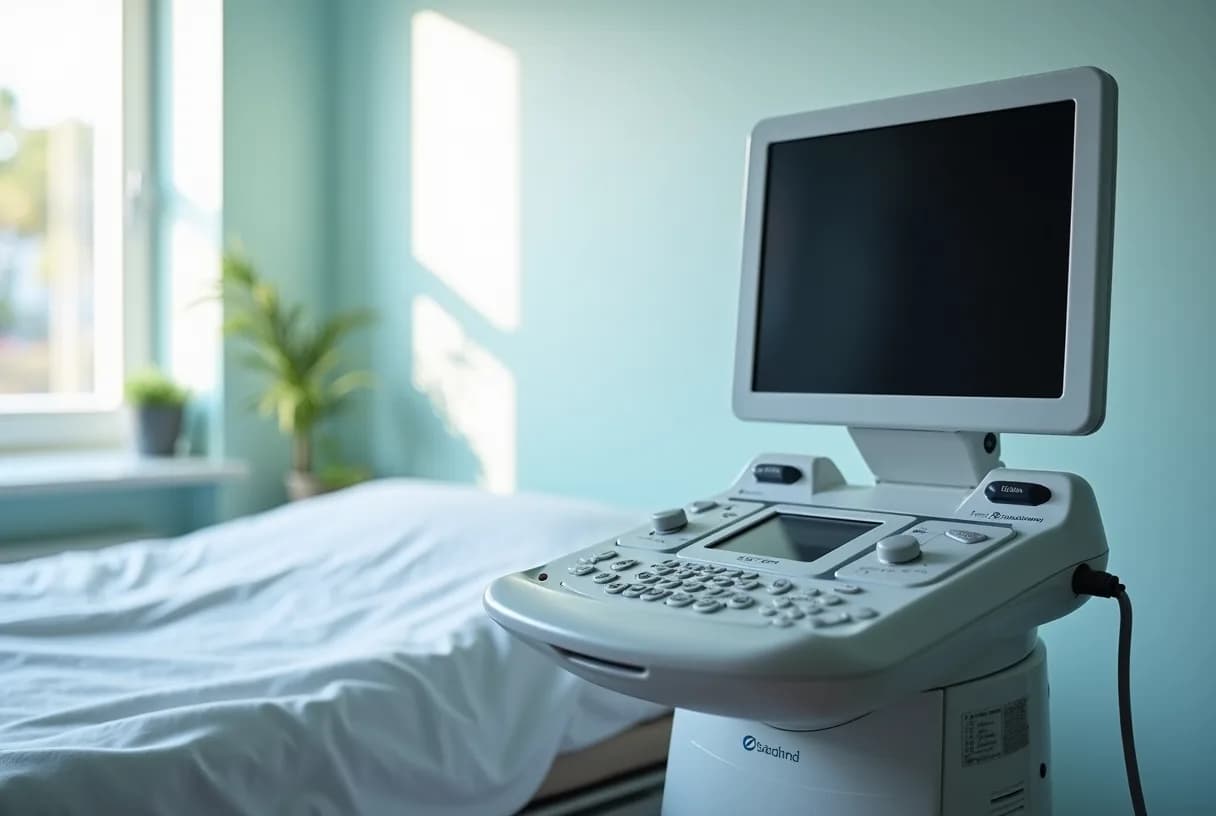
You undress from the waist down and lie on your back on a couch with a sheet over you, usually with your knees bent and feet apart, sometimes with your hips raised on a cushion so the probe angles better. The probe is roughly the width of two fingers — about 2–3 cm across — and only the tip goes in, around 5–8 cm. It is covered with a disposable sheath (tell them if you have a latex allergy) and coated with gel. The sonographer angles and rotates it to sweep across the uterus and each ovary; they may press on your lower abdomen with their other hand to bring an ovary into view, which can feel like period-type pressure.
It takes about 10–20 minutes. There is no radiation. It should feel like pressure and movement, not sharp pain. You are usually asked to empty your bladder first (the opposite of an abdominal scan, which needs a full one).
Things nobody tells you, but that are true almost everywhere:
- You can ask to insert the probe yourself. Many sonographers offer this; if they don't, ask.
- You are entitled to a chaperone, whatever the sex of the person scanning you.
- You can say stop, at any point, and it stops.
- You can be scanned on your period — take a tampon out first; a little bleeding does not ruin the image.
- If you have vaginal dryness or genitourinary syndrome of menopause, say so before they start. Extra gel, a smaller probe and a slower approach make a real difference, and if scans have hurt before it is worth asking your prescriber, well ahead of the appointment, whether treating the dryness first is appropriate for you.
- Light spotting for a day afterwards is common and not a sign anything went wrong.
- If penetration is impossible or intolerable — vaginismus, severe atrophy, no history of vaginal penetration — a transabdominal or transrectal scan is an alternative. It is a legitimate request, not a failure.
What can it see — and what can't it?
| Sees well | Sees partly / unreliably | Cannot see |
|---|---|---|
| Endometrial thickness; polyps and focal lining lesions | Adenomyosis (features are suggestive, not definitive) | Whether any tissue is cancerous — that needs a biopsy |
| Fibroids: number, size and — crucially — location (submucosal, intramural, subserosal) | Deep endometriosis nodules (only in expert hands, with a dedicated protocol) | Superficial peritoneal endometriosis — invisible on ultrasound |
| Ovarian cysts: simple vs complex, solid areas, septations, blood flow | Ovaries after menopause, or when a large fibroid or bowel gas blocks the view | Normal fallopian tubes; whether tubes are open (that needs HyCoSy or an HSG) |
| IUD position; free fluid in the pelvis; ectopic pregnancy signs | Endometrium distorted by fibroids, previous surgery or an axial uterus | Early ovarian cancer in a woman without symptoms |
Two consequences of that table matter enormously and are routinely missed in the consulting room. First: a normal scan does not rule out endometriosis. Women are told "your scan was clear" and sent away, when standard ultrasound simply cannot see the commonest form of the disease. If your symptoms fit, a normal scan is not the end of the road — see endometriosis and pelvic pain. Second: ultrasound is not a cancer screening test. Large randomised trials of annual TVUS in healthy women found no reduction in ovarian cancer deaths while producing false alarms and operations on benign cysts, and the US Preventive Services Task Force recommends against screening women at average risk. There is no screening programme for endometrial cancer either: a smear (Pap) test samples the cervix, not the womb lining, so a normal smear tells you nothing about your endometrium. Womb cancer is found by investigating symptoms — above all, bleeding.
What does endometrial thickness mean?
This is the number everyone Googles at 2 a.m., and its meaning depends entirely on who you are.
| Situation | What the measurement means |
|---|---|
| Postmenopausal, bleeding, not on hormone therapy | The one setting where the number is genuinely decisive. A thin lining — commonly a 4 mm threshold — makes endometrial cancer very unlikely (the residual risk is well under 1%) and may reasonably avoid a biopsy after a single episode. Thicker than the threshold, or a focal lesion, means the lining must be sampled. |
| Postmenopausal, bleeding, on hormone therapy or tamoxifen | Thickness thresholds are unreliable. Sequential HRT thickens the lining cyclically; tamoxifen produces sub-endometrial cysts that mimic pathology. Bleeding here still needs assessment — usually tissue sampling or hysteroscopy rather than trusting a millimetre reading. Never adjust your own HRT in response to a scan; that is a conversation with your prescriber. |
| Postmenopausal, no bleeding, thick lining found incidentally | A different question with a much weaker evidence base. The 4 mm rule does not apply, and most incidentally thickened linings are benign. Management is individualised on risk factors (obesity, diabetes, tamoxifen, Lynch syndrome, unopposed oestrogen) — not automatic biopsy. |
| Premenopausal or perimenopausal | Thickness is expected to change across the cycle — a few millimetres just after a period, over 10 mm in the second half. There is no cancer cut-off. That is why endometrial scans are best timed to days 5–10 of the cycle, and why bleeding problems here are assessed on pattern, risk factors and biopsy, not on thickness alone. |
Does a thin lining mean I'm in the clear?
No — and this is the most important sentence on this page. A thin endometrium in a bleeding postmenopausal woman makes cancer very unlikely at that moment. It does not make it impossible.
Three reasons the reassurance has limits:
- Some endometrial cancers grow on a thin, atrophic-looking lining. The aggressive type II cancers — serous and clear cell — are the ones most likely to slip past a thickness-based rule. They are less common but more dangerous.
- The scan can be technically inadequate and still be reported with a number. Fibroids, previous surgery, an angled uterus or a difficult view can all make the lining impossible to measure properly. "Endometrium not adequately visualised" is not a normal result — it means the question is unanswered and sampling is needed.
- Thickness is a snapshot; bleeding is a process. A reassuring measurement on Tuesday says nothing about what causes bleeding in three months' time.
So the rule that professional guidance actually applies is this: a thin lining can justify not doing a biopsy after a single episode of postmenopausal bleeding — but if the bleeding persists or recurs, the case reopens, regardless of how good the scan looked. That means going back, and asking for tissue: an endometrial biopsy (a pipelle, done in clinic), a saline infusion sonohysterogram, or hysteroscopy with directed sampling. If you take one thing from this article, take that. Women are lost to a "normal scan" they were told about once and never revisited.
What happens after the scan?
The sonographer usually cannot give you a diagnosis on the spot; a radiologist or your gynaecologist interprets and reports. Typical next steps:
- Thickened lining or focal lesion + bleeding → endometrial biopsy, or hysteroscopy (a camera inside the uterus) if the biopsy is non-diagnostic or a polyp needs removing.
- Suspected polyp or submucosal fibroid → saline infusion sonohysterography, which floats the cavity open with fluid and shows the lining in far more detail, or hysteroscopy.
- Simple ovarian cyst → often just a repeat scan in a few months; most resolve. See ovarian cysts.
- Complex or solid ovarian mass → a risk score, a CA-125 blood test, and referral to a gynaecological oncology service if the risk is raised.
- Normal scan but ongoing symptoms → the symptoms still need explaining. A normal scan is a finding, not a verdict.
Ask for a copy of the report, not just a phone call. If you're building a case for being taken seriously, our doctor report tool can help you turn a symptom diary into something a 10-minute appointment can act on, and gynecologic health covers the wider picture.
When to see a doctor
Book an urgent appointment — do not wait to see if it settles — if you have:
- Any vaginal bleeding or spotting after menopause (12 months with no period). One episode counts. On HRT, unexpected or persistent bleeding also needs to be checked with your prescriber — don't stop or change the dose yourself.
- Watery, pink or blood-stained discharge after menopause, even without frank bleeding. It is most often caused by a thin, atrophic lining or an infection, but it warrants the same investigation as bleeding, because endometrial cancer can present as discharge with no visible blood.
- Bleeding that returns after a reassuring scan. Go back. Say the words: "the bleeding has continued and I would like the lining sampled."
- Bleeding after sex, at any age.
- Persistent bloating, feeling full quickly, loss of appetite, or new urinary urgency occurring most days for three weeks or more — the pattern that should prompt an ovarian cancer assessment.
- Periods that are suddenly much heavier, or bleeding between periods, in perimenopause.
Go to urgent or emergency care for sudden severe one-sided pelvic pain, especially with nausea or vomiting — this can mean a cyst has ruptured or an ovary has twisted (torsion), and it is time-critical.
One more thing worth saying out loud, because it is common: if you have reported bleeding and been told it's "just perimenopause" or "just stress" without an examination or a scan, that is not an assessment. Ask, in writing if you have to, why a transvaginal ultrasound is not indicated. You are allowed to be the one who insists.
]]>